spatialHeatmap
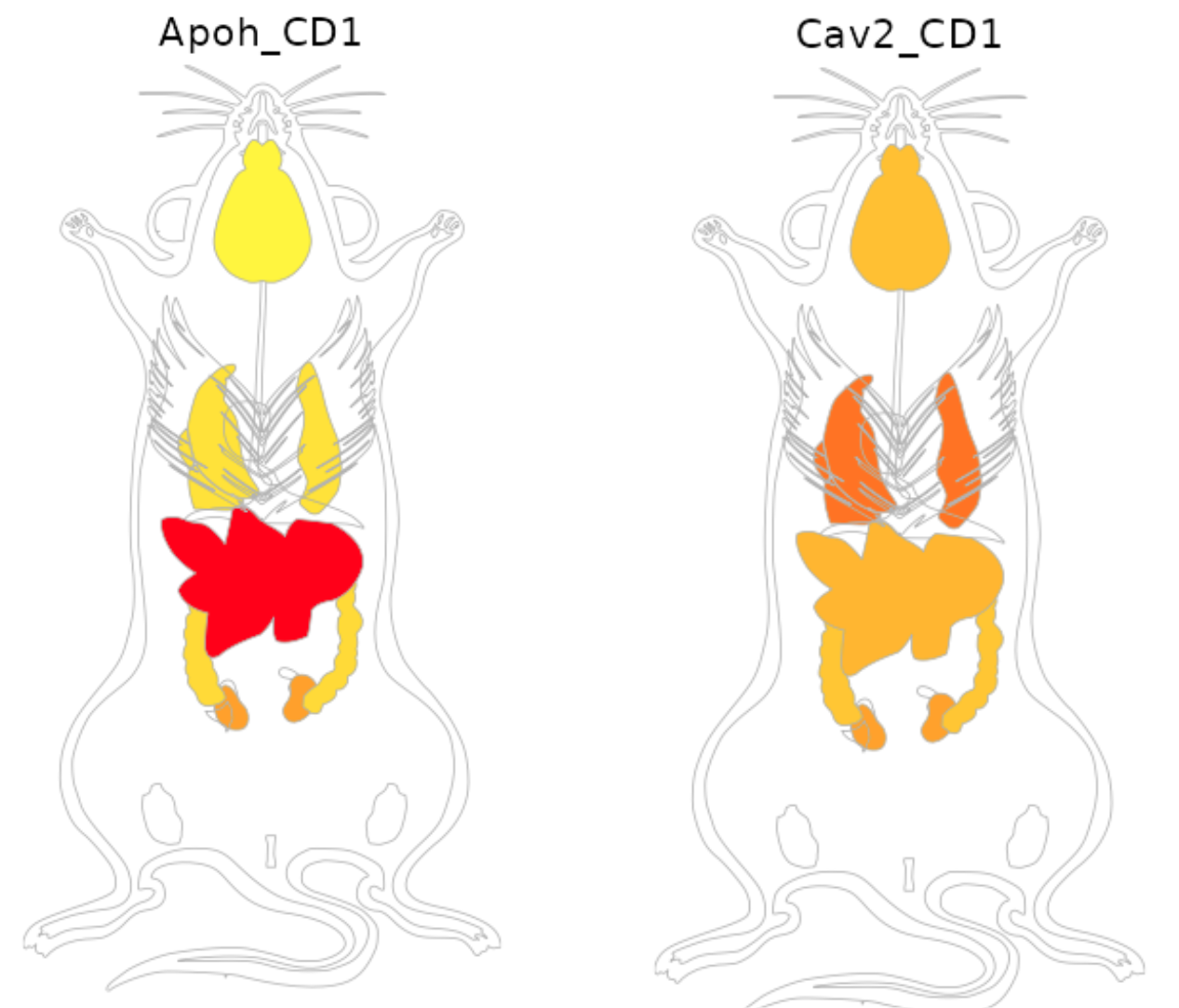
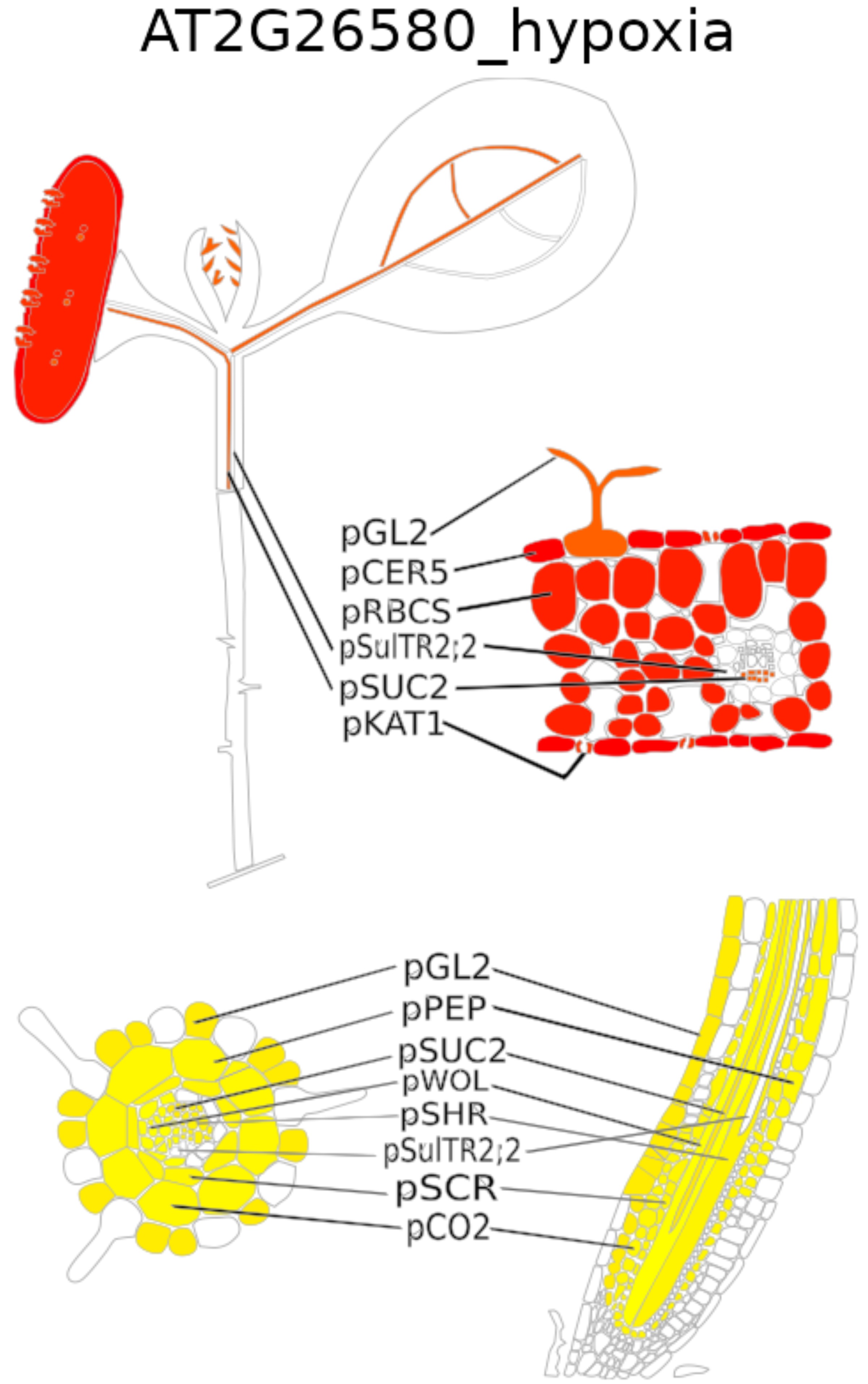
The spatialHeatmap software visualizes spatial bulk and single cell assays in anatomical images.
Getting started: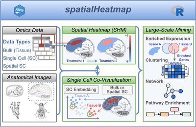
Functionalities
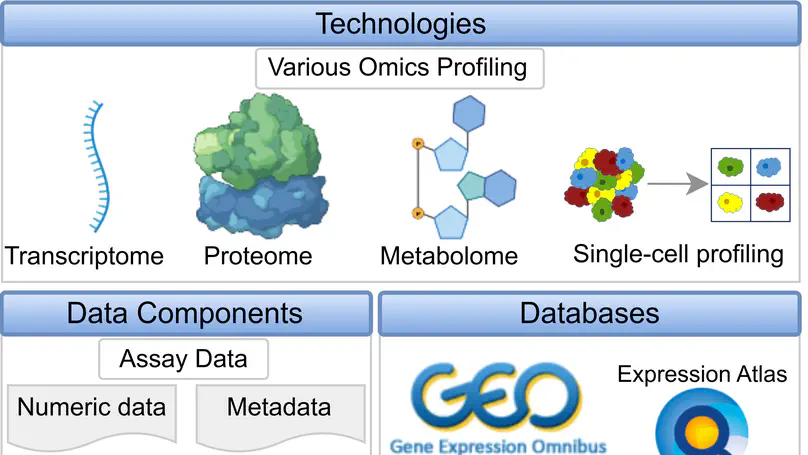
The software supports anatomical images from public repositories (e.g. EBI anatomogram) or those created by users. In general any type of image can be used as long as it can be provided in SVG (Scalable Vector Graphics) format and the corresponding spatial features, such as organs, tissues, cellular compartments, are annotated with unique identifiers.
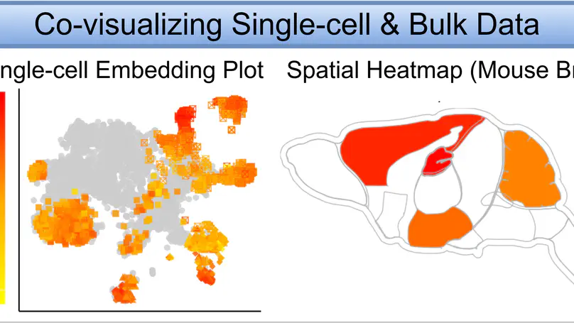
The spatialHeatmap software provides novel functionalities to integrate tissue and single-cell data by visualizing them in composite plots that combine SHMs with embedding plots of high-dimensional data. Identical colors indicate matching components between SHM and embedding plots.

To appeal to both non-expert and computational users, spatialHeatmap provides a graphical and command-line interface, respectively. It is distributed as a free, open-source Bioconductor package (https://bioconductor.org/packages/spatialHeatmap) that users can install on personal computers, shared servers, or cloud systems.


